The Shaner Lab
UCSD Departments of Neurosciencesand Pharmacology
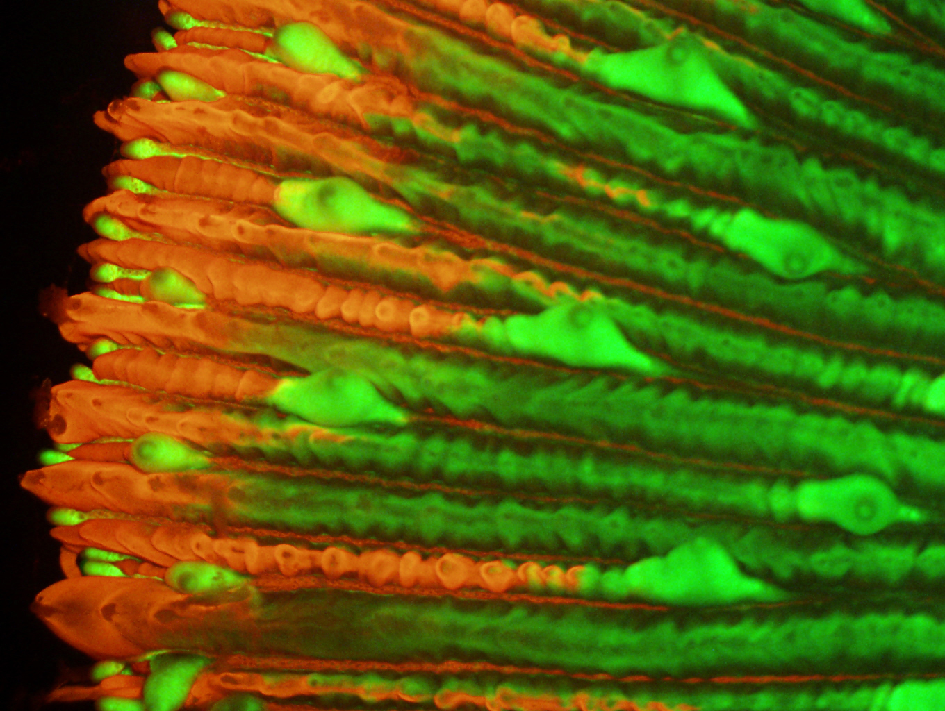
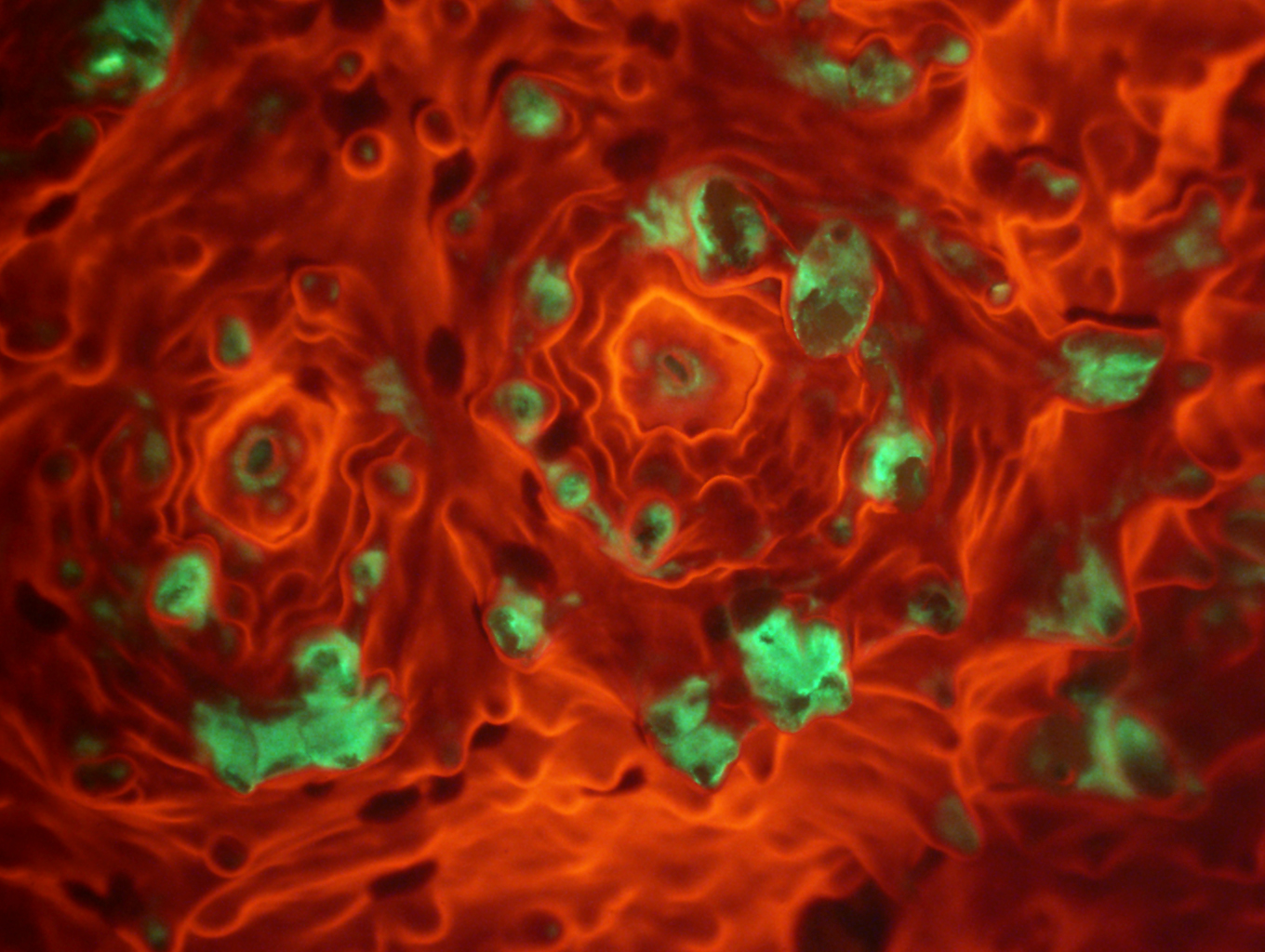
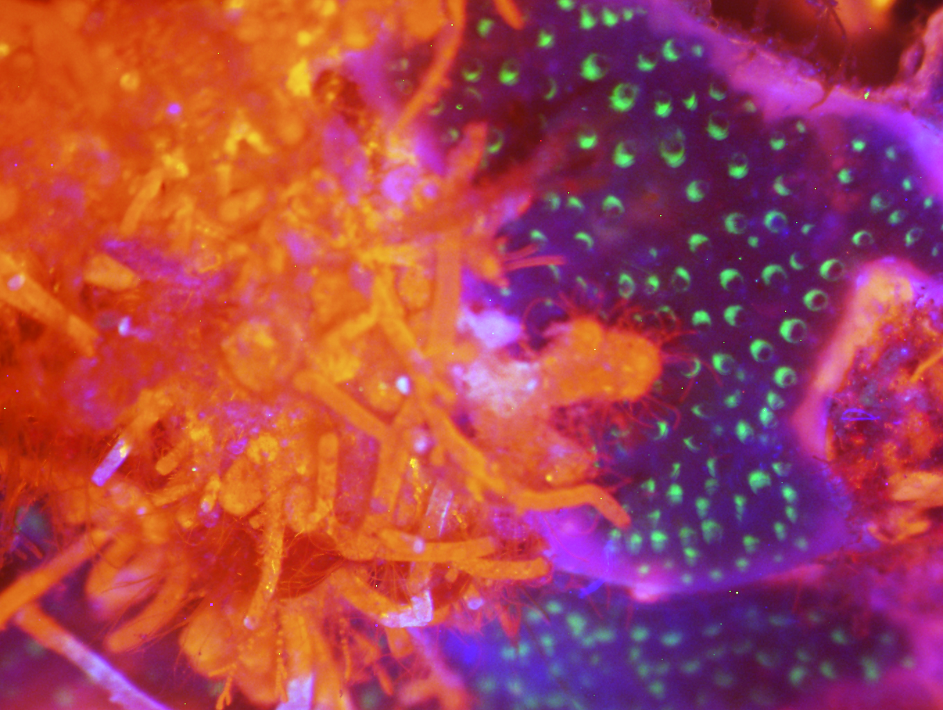
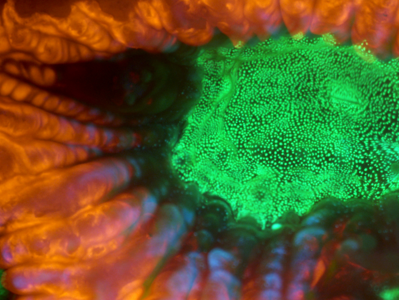
We adapt the chemistry of naturally occurring fluorescent and bioluminescent proteins to build optical probes that report and control cellular activity with high precision.
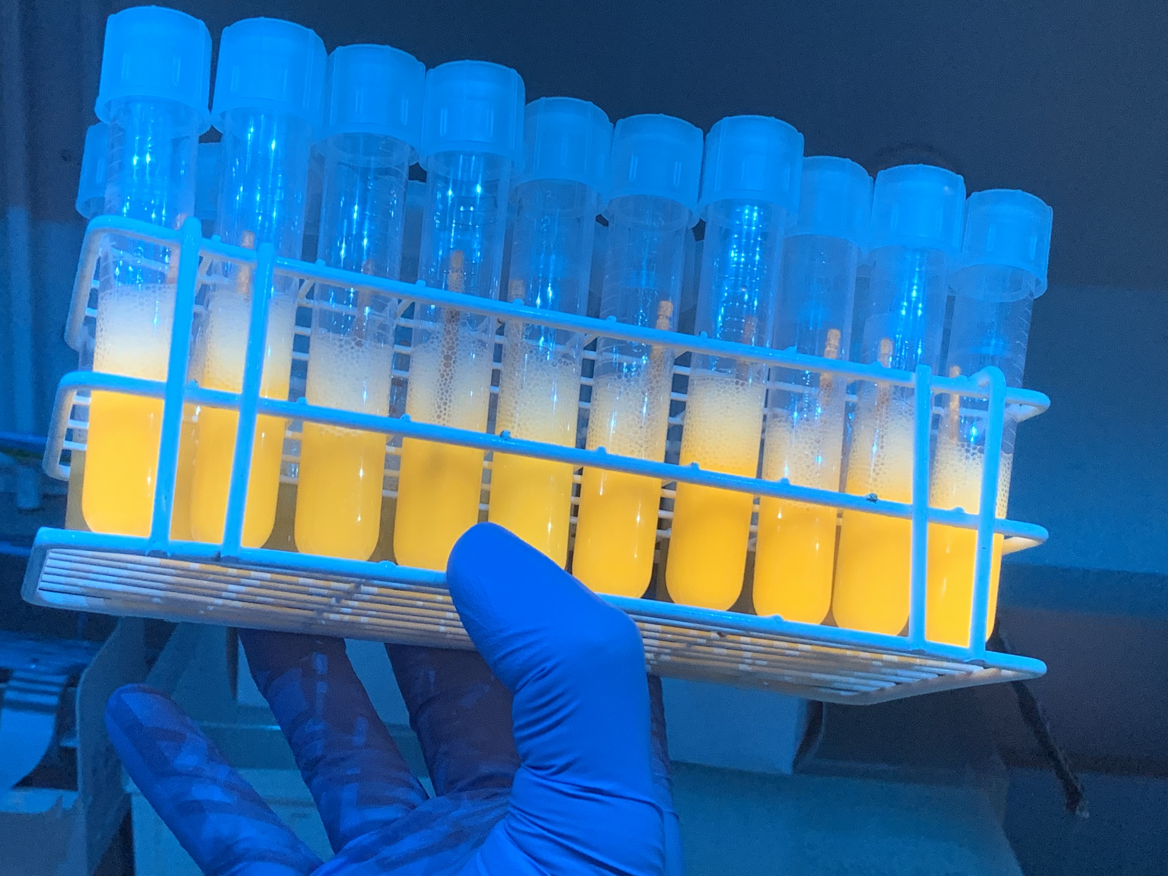
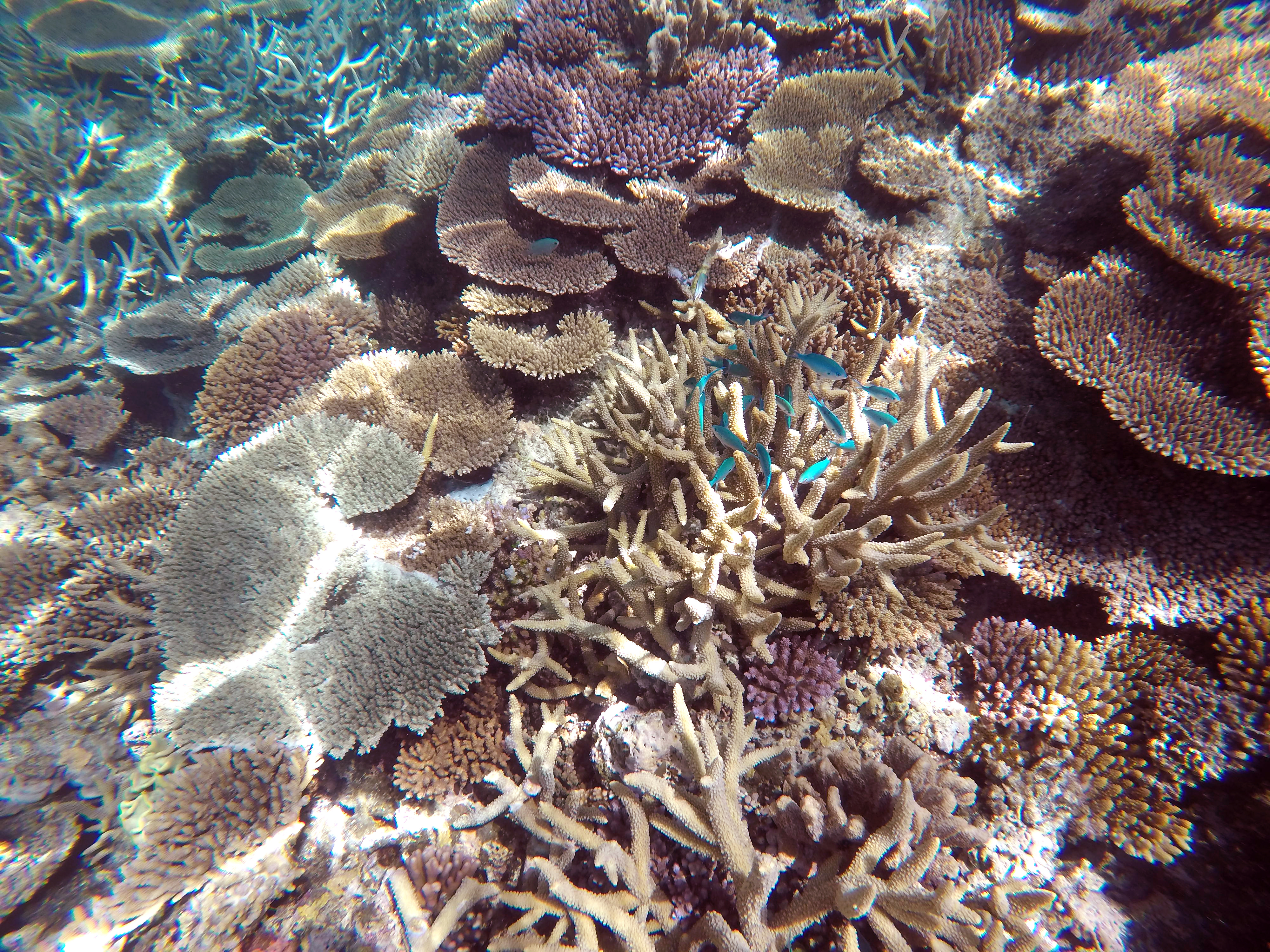
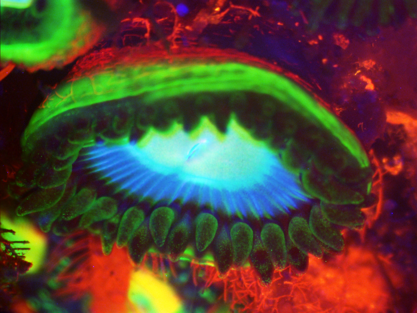
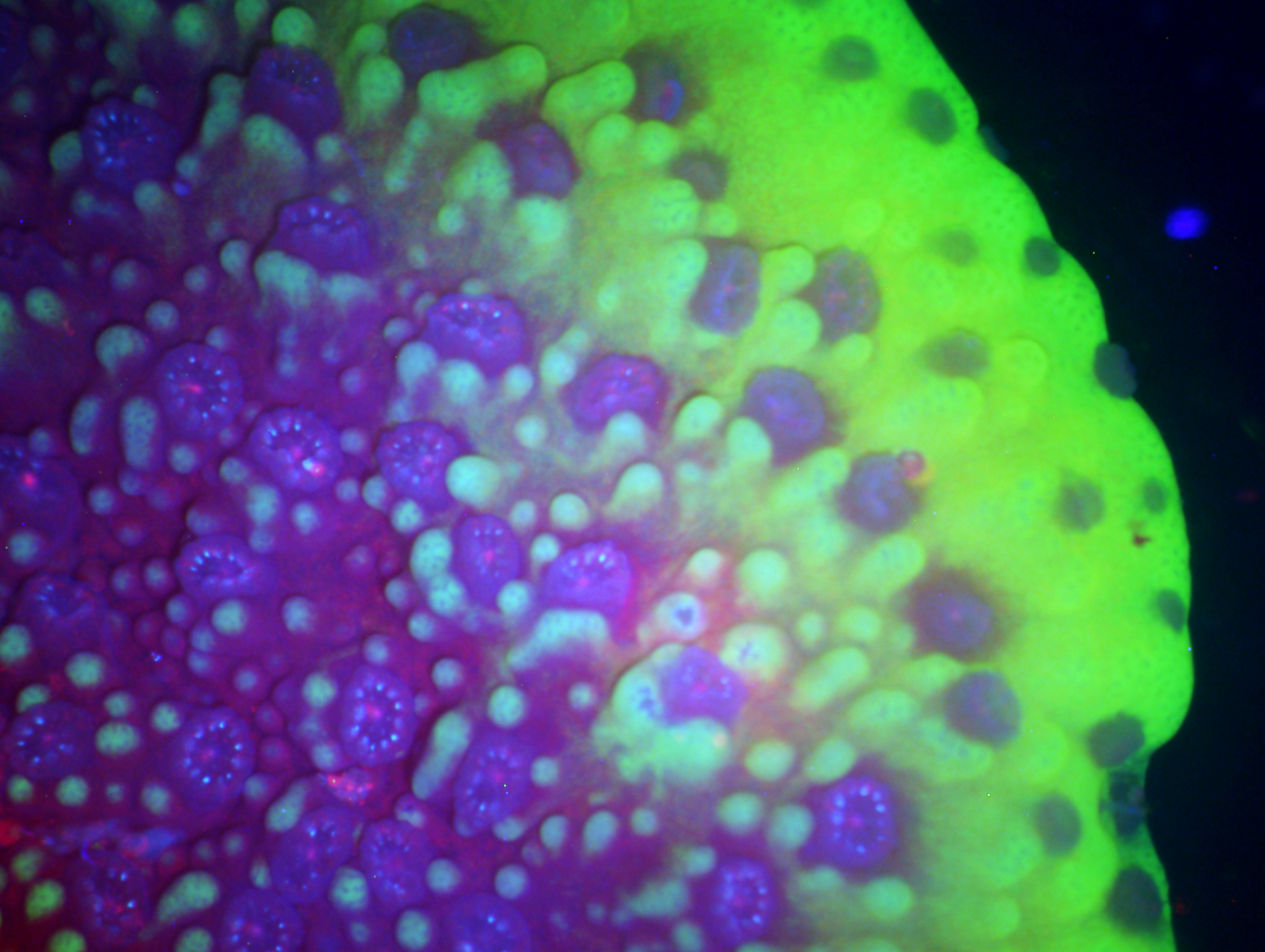
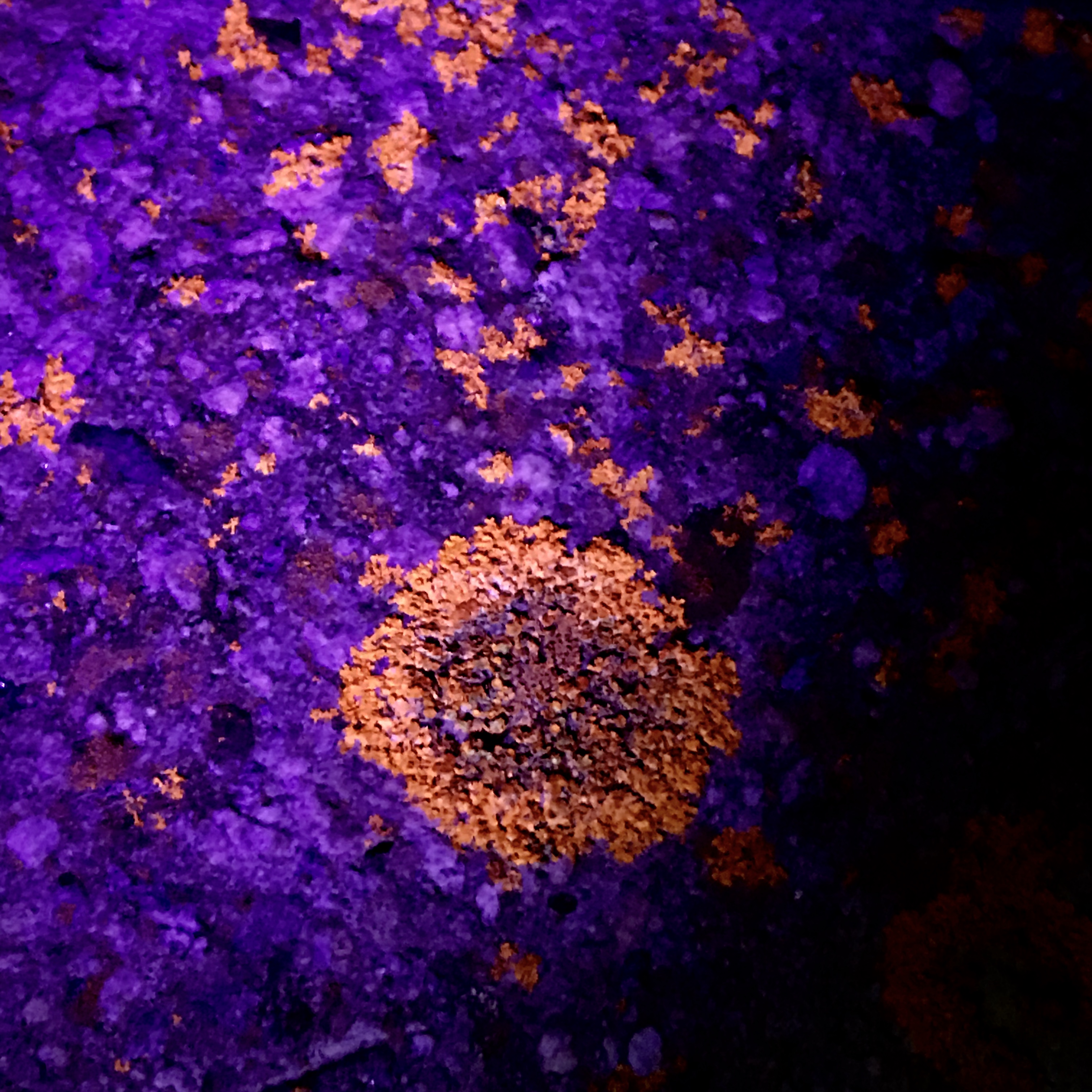
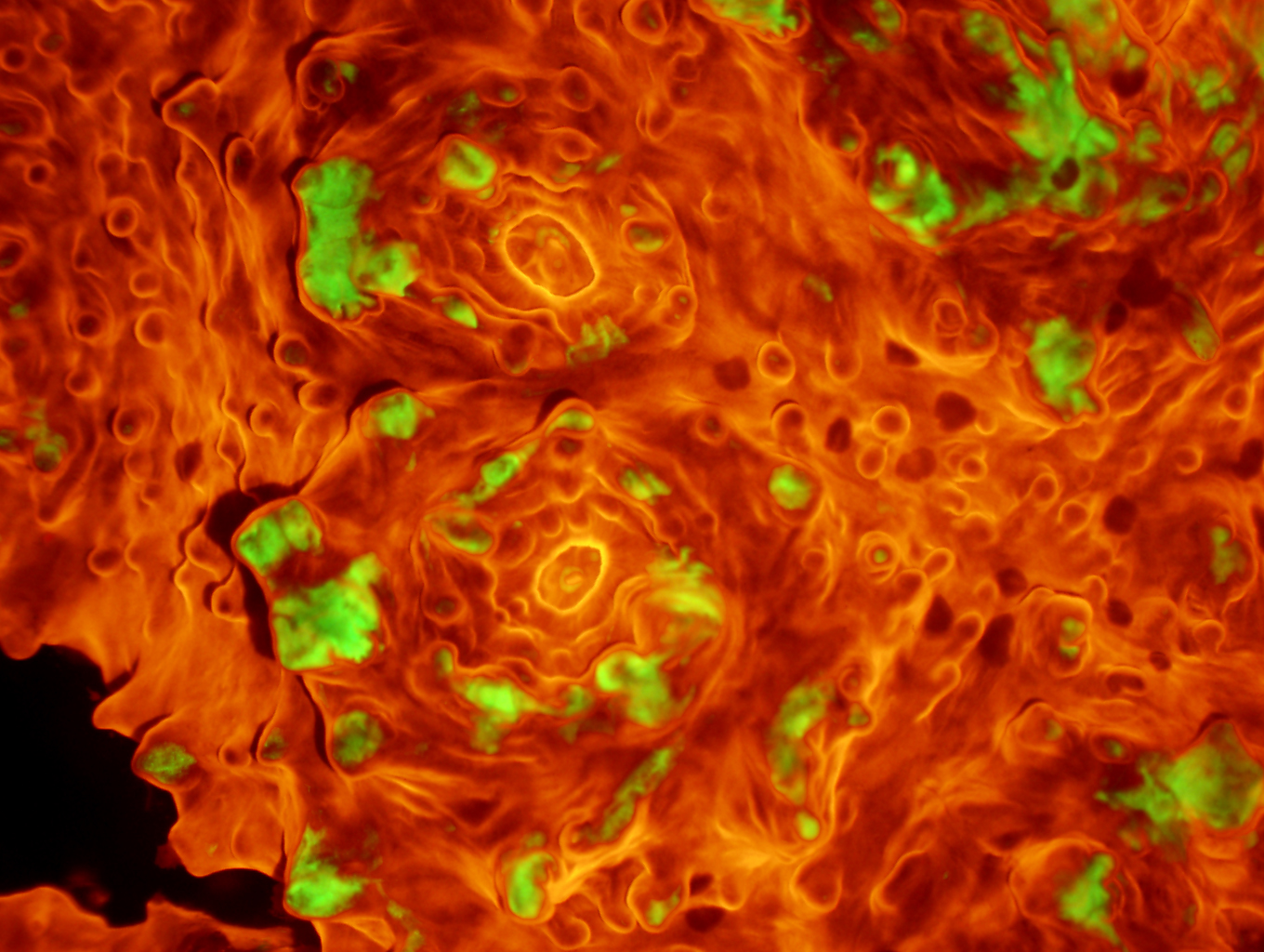
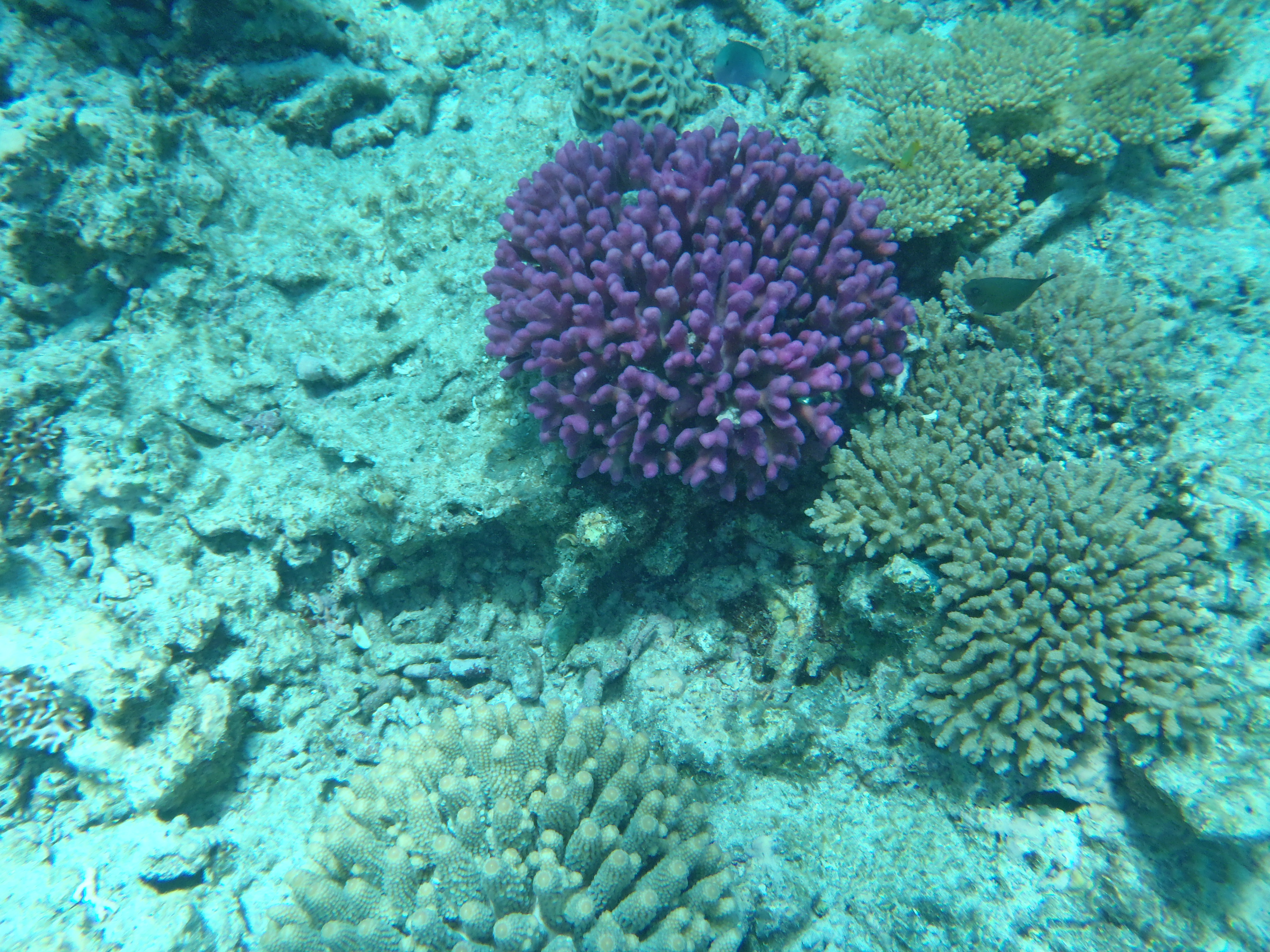
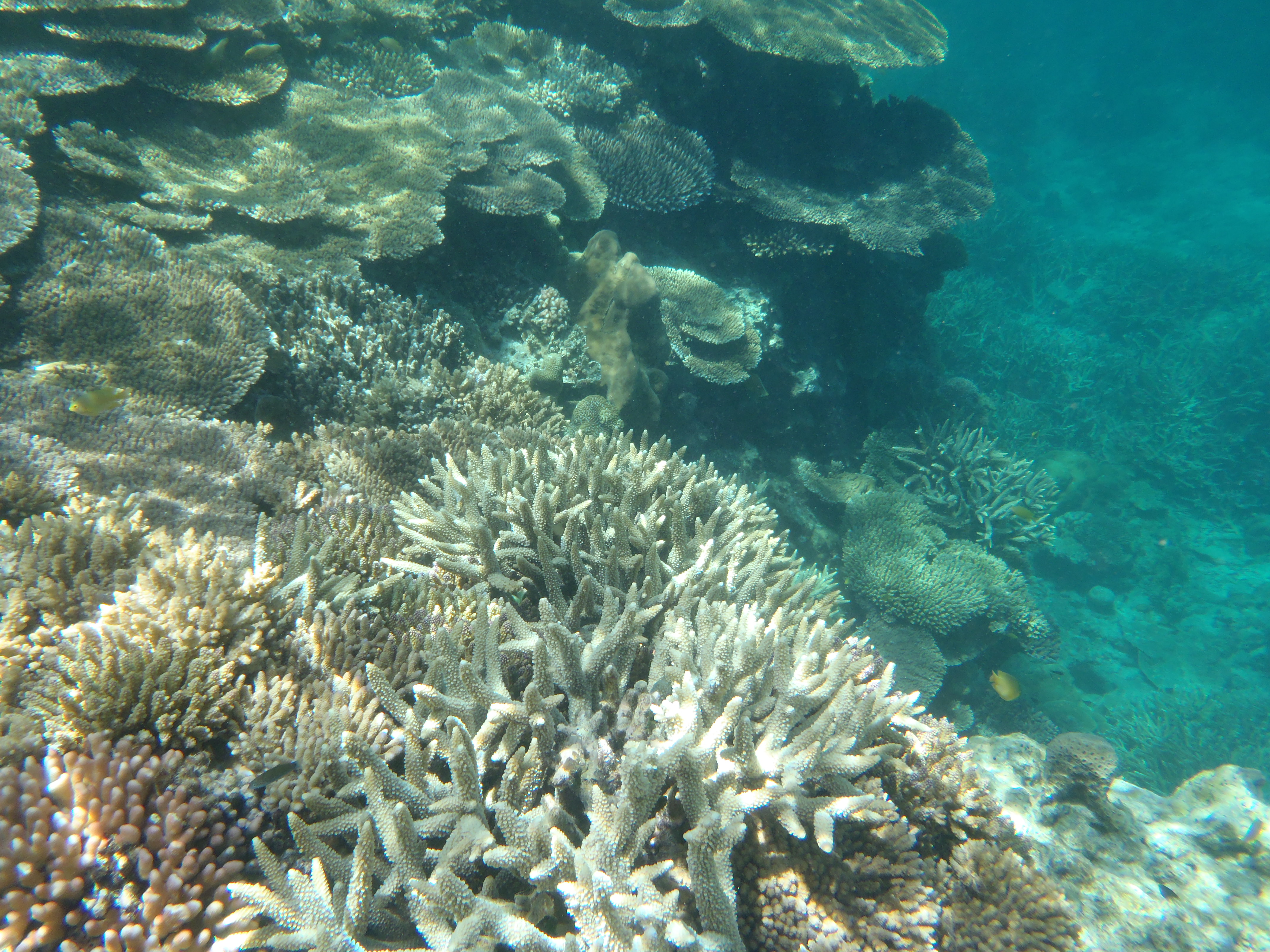
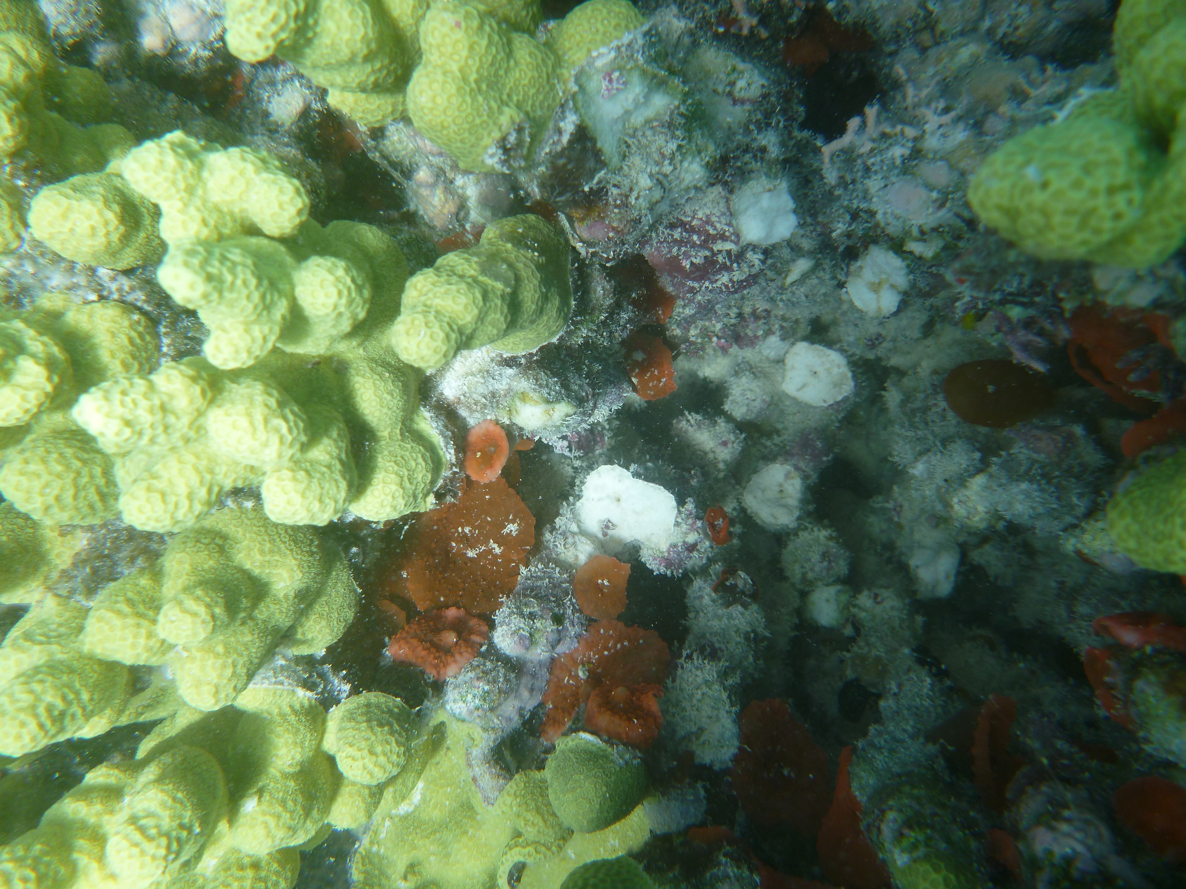
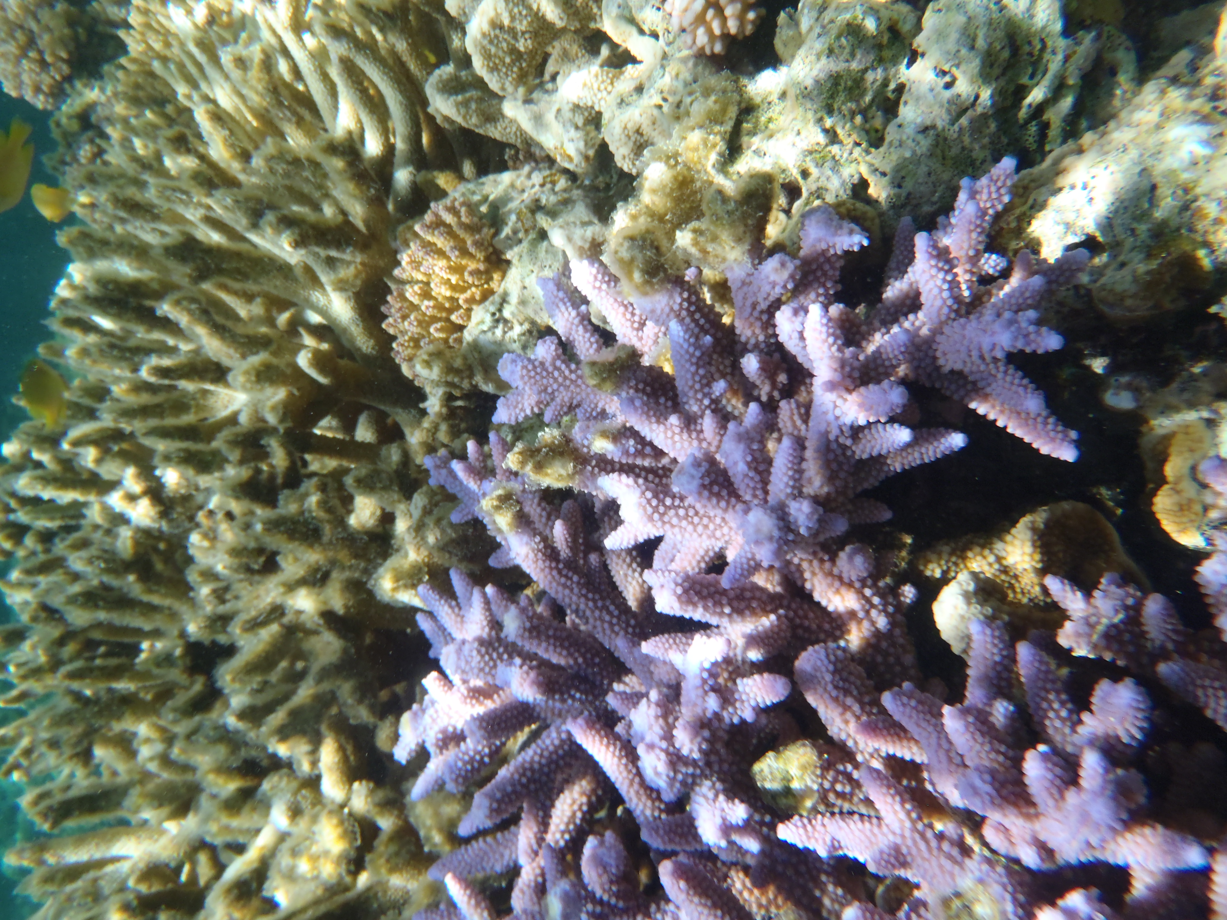
Our field collections and transcriptome mining of corals, jellyfish, and other marine organisms yield novel protein scaffolds for imaging and biosensing.
By fusing luciferases to light-sensitive ion channels, we create luminopsins that enable chemogenetic delivery of light for non-invasive neural control.
This engineered indicator detects neural and glial calcium signals in vivo without excitation light, enabling long-term, stable recording in freely behaving animals.
The mGold2 family resists photobleaching during extended imaging, addressing a long-standing limitation of YFPs for quantitative microscopy.
Luciferin-gated fluorescent reporters integrate transient signals over time, converting dynamic activity into stable readouts for later analysis.
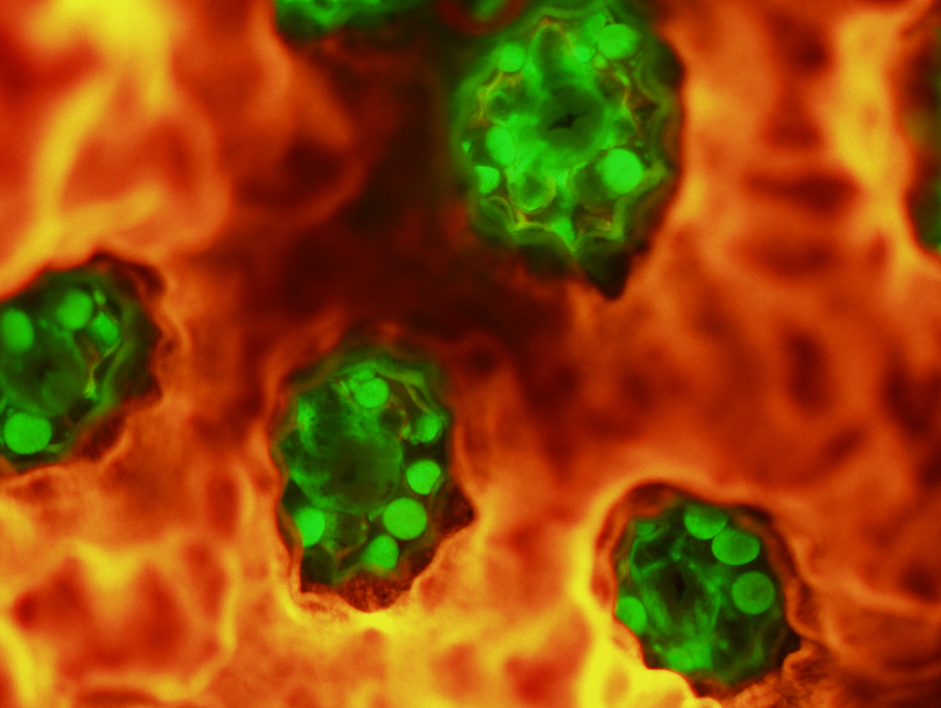
Our optical tools allow continuous monitoring of immune cells, metastatic cancer cells, and other motile populations in vitro and in vivo.
We provide consultation, assay development, and probe design to help academic and industry researchers integrate biosensors into their projects.
We develop peptide and transmitter indicators to study how marine nervous systems respond to warming, acidification, and other stressors.
Bioluminescent sensors track CAR T-cell activation patterns, providing insight into mechanisms limiting solid tumor immunotherapy.
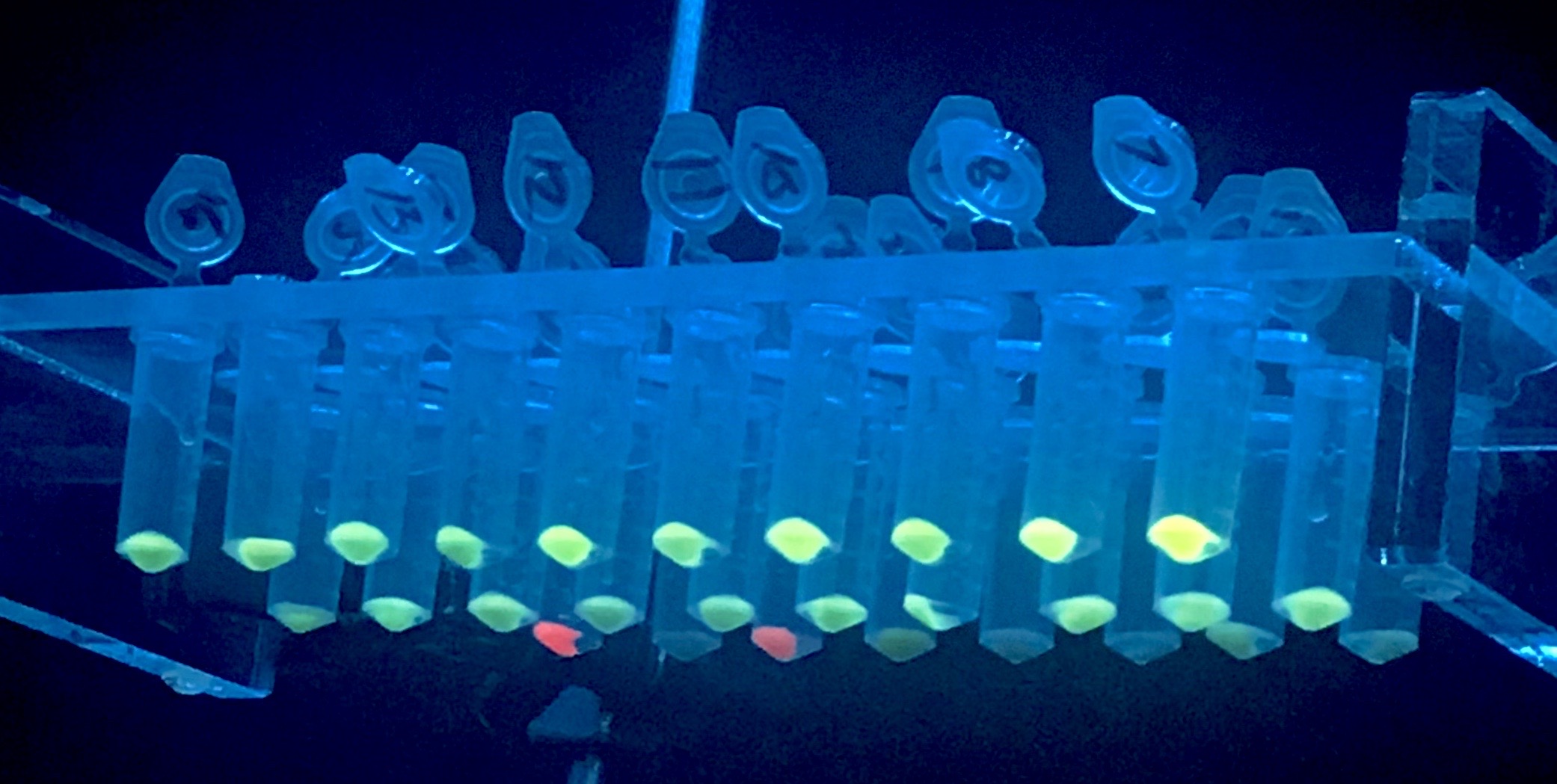
Distinct bioluminescent chemistries enable simultaneous optical control and readout of multiple biological processes without spectral crosstalk.
We demonstrate functional connectivity between neurons built entirely from bioluminescent sources and optogenetic actuators.
Our group distributes probes and protocols without restriction, fostering rapid validation and application across diverse systems.
We tune chromophore environments to optimize brightness, photostability, and spectral properties for demanding imaging tasks.
Bioluminescence enables quantitative in vivo imaging using simple optics, lowering barriers for under-resourced laboratories.
Transcriptomic data mining and directed evolution link novel sequences to functional probes with tailored performance.
Our projects bring together neuroscientists, marine biologists, chemists, engineers, and clinicians to apply optical tools to new questions.
Undergraduate, graduate, and postdoctoral researchers learn optical probe engineering and imaging through hands-on mentorship.
We design sensors to be compatible with standard microscopes, enabling use in labs without specialized equipment.
Our probes transform molecular events into photons, making invisible processes visible and quantifiable.